Three-Dimensional Ultra-High Frequency Ultrasound Facilitates Image Processing to Visualize Microstructural Changes of Hair Follicles and Detects Distinct Disease Phases of Alopecia Areata
April 2023
in “
Journal of Investigative Dermatology
”
TLDR 3D ultrasound can detect hair follicle changes and disease phases in alopecia areata.
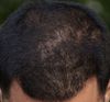
The study introduces a three-dimensional ultra-high frequency ultrasound (3D-uHFUS) as a novel non-invasive diagnostic tool for alopecia areata (AA), overcoming limitations of traditional methods by capturing both transverse and vertical images of hair follicles. Involving 18 AA patients, the 3D-uHFUS successfully visualized hair cycle status and inflammation severity in 15 cases, aligning with clinical records. Unique pathological signs, such as hyperechogenic ovoid structures (HOSs) and inverse triangular hypoechogenicity (ITH), were identified, with HOSs linked to poor prognosis and ITH to acute AA phases. These findings, not detectable by conventional tools, suggest 3D-uHFUS's potential for early prediction of AA's clinical course and treatment response.