Fundus Changes in Systemic Lupus Erythematosus
April 2026
in “
The National Medical Journal of India
”
TLDR Regular eye exams are important for detecting serious complications in lupus patients.
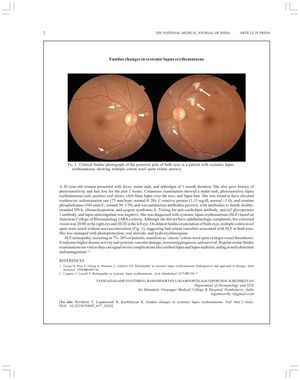
A 42-year-old woman with systemic lupus erythematosus (SLE) presented with symptoms including fever, malar rash, arthralgia, photosensitivity, and hair loss. Despite no ophthalmologic complaints, a fundus examination revealed multiple cotton wool spots, indicating retinal vasculitis associated with SLE. This condition, known as SLE retinopathy, occurs in 7%-26% of patients and is linked to higher disease activity and systemic vascular damage, which can worsen prognosis and survival. The patient was treated with photoprotection, oral steroids, and hydroxychloroquine. Regular ocular fundus examinations are crucial for early detection and management of severe complications such as cerebral lupus and lupus nephritis.