Nevus Sebaceous: Histopathological Examination
January 2025
in “
Indian Journal of Dermatopathology and Diagnostic Dermatology
”
nevus sebaceous epidermal acanthosis papillomatosis hair follicle density sebaceous glands apocrine glands basaloid arrangement follicular germinative cells adnexal epithelial proliferation sebaceous hyperplasia immature follicular units basaloid adnexal elements basal cell carcinoma trichoblastoma syringocystadenoma papilliferum follicular hamartoma epidermal nevus
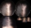
TLDR Nevus sebaceous is identified by unique skin changes, including thickened skin, fewer hair follicles, and many sebaceous glands.
The histopathological examination of nevus sebaceous (NS) reveals distinct features such as focal epidermal acanthosis, papillomatosis, and a significant reduction in hair follicle density, with remaining follicles being rudimentary and some filled with keratin. The dermis contains numerous mature sebaceous glands, often lacking visible ducts, and scarce apocrine glands located beneath the sebaceous units. A basaloid arrangement of follicular germinative cells indicates adnexal epithelial proliferation. These characteristics—sebaceous hyperplasia, immature follicular units, and basaloid adnexal elements—are indicative of NS and help differentiate it from other conditions like sebaceous hyperplasia, basal cell carcinoma, trichoblastoma, syringocystadenoma papilliferum, follicular hamartoma, and epidermal nevus. The integration of epidermal, sebaceous, follicular, and apocrine alterations is crucial for a definitive diagnosis of NS.