 3 citations
,
October 2015 in “International Journal of Dermatology”
3 citations
,
October 2015 in “International Journal of Dermatology” Normal hair density and diameter ratios were established to help diagnose adult hair loss.
8 citations
,
June 1981 in “Clinica Chimica Acta”  February 2026 in “Zenodo (CERN European Organization for Nuclear Research)”
 Trichoscopy is important for planning hair restoration in burn-related hair loss.
 9 citations
,
January 2019 in “Skin appendage disorders”
9 citations
,
January 2019 in “Skin appendage disorders” Researchers found a new sign of fungal infection in some patients with Seborrheic Dermatitis.
 16 citations
,
January 2010 in “American Journal of Neuroradiology”
16 citations
,
January 2010 in “American Journal of Neuroradiology” Specific brain and bone imaging findings can help diagnose Trichothiodystrophy.
OCT can effectively diagnose different stages of hair loss non-invasively.
 January 2023 in “Asian Journal of Pediatric Research”
 Trichoscopy is crucial for diagnosing and assessing alopecia areata in children.
 8 citations
,
July 2014 in “Anais Brasileiros de Dermatologia”
8 citations
,
July 2014 in “Anais Brasileiros de Dermatologia” A man's scalp condition was misidentified as hair loss dots but was actually a common follicular disorder.
1 citations
,
May 2024 in “Skin Appendage Disorders” Early diagnosis with trichoscopy can improve management and quality of life for CCCA patients.
11 citations
,
July 2019 in “Pediatric dermatology” Children with alopecia areata have different trichoscopic features than adults, including more empty follicular openings and pigtail hairs.
 September 2022 in “bioRxiv (Cold Spring Harbor Laboratory)”
 A new staining method shows a special area in the hair's skin layer with lots of proteoglycans.
 October 1990 in “Archives of Dermatology”
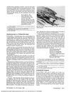
 Hair loss and growth can be accurately measured using computer-assisted counting.
July 2025 in “Indian Dermatology Online Journal” Trichotillomania incognita can mimic hair loss patterns and requires careful diagnosis to avoid misdiagnosis.
May 1988 in “Journal of Forensic Sciences” A new method accurately determines hair blood type and can be used on dust samples.
 January 2024 in “İstanbul Kuzey Klinikleri”
 Low ferritin levels are linked to increased hair loss; no link found between vitamin B12, TSH levels, and hair loss.
Non-invasive methods can effectively diagnose and manage alopecia areata.
11 citations
,
January 2017 in “Skin Appendage Disorders” Use lower concentrations of triamcinolone acetonide to reduce steroid-induced scalp atrophy.
7 citations
,
January 2007 in “PubMed” Videodermoscopy is a helpful, non-invasive tool for accurately diagnosing hair and scalp disorders.
27 citations
,
April 1978 in “Journal of Forensic Sciences” Enzyme typing can reliably characterize human hair.
13 citations
,
January 1985 in “International Journal of Dermatology” The document found differences in lesion distribution and microscopic features among trichostasis spinulosa and similar skin conditions.
A rapid screening method using trichoscopy and clinical data can improve diagnosis and treatment of tinea capitis.
January 2026 in “British Journal of Dermatology” The ATP assay can measure skin microbiome changes and recovery, with ethanol-treated skin taking longer to recover than tape-stripped skin.
 March 2021 in “CRC Press eBooks”
 Trichoscopy helps tell apart Lichen planopilaris and Frontal fibrosing alopecia from other hair loss conditions.
May 2005 in “Hair transplant forum international” Trichotemnomania causes hair loss due to compulsive hair cutting or shaving.
 40 citations
,
December 2014 in “Indian Journal of Dermatology”
40 citations
,
December 2014 in “Indian Journal of Dermatology” Two new signs, 'signet ring vessels' and 'hidden hairs,' help tell apart scalp psoriasis and seborrheic dermatitis.
26 citations
,
January 1983 in “PubMed” Trichothiodystrophy involves brittle hair due to low sulfur amino acids, not a transport defect.
January 2006 in “The Year book of dermatology”  February 2026 in “Frontiers in Medicine”
 Trichoscopy can help diagnose early congenital syphilis in newborns.
 42 citations
,
January 2011 in “Journal of Biomedical Optics”
42 citations
,
January 2011 in “Journal of Biomedical Optics” Infrared and Raman imaging can non-destructively analyze hair structure and help diagnose hair conditions.
Trichoepithelioma and desmoplastic trichoepithelioma have distinct features that can be identified using reflectance confocal microscopy.