 17 citations
,
December 2014 in “Cell Stem Cell”
17 citations
,
December 2014 in “Cell Stem Cell” Intravital imaging helps us better understand stem cells in their natural environment and could improve knowledge of organ regeneration and cancer development.
21 citations
,
July 2004 in “Apmis” Fluorescent proteins help visualize and understand tumor blood vessel growth.
 9 citations
,
January 2011 in “Skin Research and Technology”
9 citations
,
January 2011 in “Skin Research and Technology” The new automatic tool accurately measures hair thickness and is reliable.
 June 2020 in “Journal of Investigative Dermatology”
 The technique effectively shows how human skin and hair cells form into ball-like structures.
9 citations
,
November 2006 in “Journal of Microscopy” Dye mainly stays on the hair surface and endocuticle.
March 2026 in “Mendeley Data” March 2026 in “Mendeley Data” 3 citations
,
October 2006 in “Journal of dermatology” X-ray microscopy can non-invasively show hair structure changes after treatments, but it's less detailed than TEM and needs improvement.
10 citations
,
January 2020 in “Royal Society Open Science” A new automated method accurately measures hair damage using microscopic images.
 30 citations
,
September 2009 in “Seminars in Cutaneous Medicine and Surgery”
30 citations
,
September 2009 in “Seminars in Cutaneous Medicine and Surgery” Dermoscopy has greatly improved the diagnosis of skin lesions and our understanding of their morphology and biology.
 18 citations
,
September 2013 in “Technology”
18 citations
,
September 2013 in “Technology” The study introduced a new imaging technology to track skin healing and bone marrow cell activity over time.
 37 citations
,
July 2016 in “Current Opinion in Cell Biology”
37 citations
,
July 2016 in “Current Opinion in Cell Biology” Live imaging has advanced our understanding of stem cell behavior and raised new research questions.
January 1994 in “Medical Entomology and Zoology” Electron microscopy helps understand skin structure and diseases.
August 2000 in “Microscopy and Microanalysis” The method successfully visualizes iodine in biological tissues.
 June 2002 in “Seminars in Cutaneous Medicine and Surgery”
 Using a microscope during hair transplants cuts damage to follicles in half and could improve hair growth.
1 citations
,
June 2012 in “OhioLink ETD Center (Ohio Library and Information Network)” A new 3-D bioreactor system improves drug screening and reduces animal testing.
January 1991 in “Acta Dermato Venereologica” A new method effectively visualizes keratin in hair without harsh chemicals.
 44 citations
,
August 2008 in “Archives of Dermatology”
44 citations
,
August 2008 in “Archives of Dermatology” Trichoscopy is a non-invasive way to diagnose hair and scalp problems without needing hair samples.
1 citations
,
January 2010 in “Biological and medical physics series” Human hair's structure and properties were studied using advanced microscopes and mechanical tests.

Polarized microscopy helps identify hair irregularities in genetic disorders.

The ProScope HR is an effective, user-friendly, and affordable tool for diagnosing hair loss.
 2 citations
,
January 2025 in “Dermatology Practical & Conceptual”
2 citations
,
January 2025 in “Dermatology Practical & Conceptual” Nailfold dermoscopy is a simple, cheap way to check blood vessel changes in COVID-19 patients.
 9 citations
,
October 2022 in “Nature Communications”
9 citations
,
October 2022 in “Nature Communications” The DiLiCre mouse model is an effective tool for precise genome editing using light.
 2 citations
,
May 2023 in “Experimental dermatology”
2 citations
,
May 2023 in “Experimental dermatology” New imaging techniques can assess and track changes in mouse acne without harm, aiding treatment choices.
 4 citations
,
May 2013 in “Dermatologic Surgery”
4 citations
,
May 2013 in “Dermatologic Surgery” Three new techniques simplify and improve the preparation of tissue samples for skin cancer surgery.
24 citations
,
January 1969 in “Archives of Dermatological Research” Hair malformations may occur due to timing issues in hair development.
1 citations
,
May 2016 in “Journal of nature and science” The method successfully imaged hair follicles under the skin using iron nanoparticles.
8 citations
,
March 2023 in “BMC Research Notes” Laser-capture microdissection effectively analyzes hair follicle microbiomes, revealing region-specific bacterial differences.
 2 citations
,
January 2020
2 citations
,
January 2020 The document describes a method for preparing hair for microscopy by embedding it in plastic, cutting it, and storing it cold before imaging.
 1 citations
,
February 2018 in “Australasian journal of dermatology”
1 citations
,
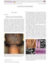
February 2018 in “Australasian journal of dermatology” Advanced imaging techniques are crucial for accurately diagnosing Monilethrix, a rare hair disorder.