January 1991 in “Acta Dermato Venereologica” A new method effectively visualizes keratin in hair without harsh chemicals.
 6 citations
,
July 2013 in “Molecular Imaging”
6 citations
,
July 2013 in “Molecular Imaging” The technique allowed noninvasive tracking of hair stem cell survival and growth, showing potential for hair loss research.
 1 citations
,
December 2014 in “Scanning”
1 citations
,
December 2014 in “Scanning” Multiphoton microscopy effectively images rabbit skin structures in detail without staining and shows differences from human skin.
 15 citations
,
July 2002 in “Clinical and Experimental Dermatology”
15 citations
,
July 2002 in “Clinical and Experimental Dermatology” Hair microscopy is useful for diagnosing certain hair loss conditions but has limitations and must be interpreted carefully.
27 citations
,
November 2012 in “Journal of Biomedical Optics” Confocal Raman microscopy can effectively study drug delivery in hair follicles using pig ear models.
 May 2012 in “The Journal of Nuclear Medicine”
 Hair stem cells were tracked in mice using a special imaging technique, showing that it's possible to monitor hair growth this way.
2 citations
,
November 2025 in “Pharmaceutics” Moderately lipophilic dyes penetrate skin deeply, while highly hydrophobic or lipophilic dyes stay on the surface.
April 2026 in “Spectrochimica Acta Part A Molecular and Biomolecular Spectroscopy” 29 citations
,
April 2011 in “Journal of Microscopy” The modified osmium method improves hair cortex staining for better visualization.
 1 citations
,
October 2024 in “Dermatology Practical & Conceptual”
1 citations
,
October 2024 in “Dermatology Practical & Conceptual” Reflectance confocal microscopy is a promising non-invasive tool for diagnosing alopecia areata incognita.
 39 citations
,
June 1982 in “The BMJ”
39 citations
,
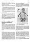
June 1982 in “The BMJ” Blood tests confirmed a baby in the womb had a CMV infection.
 3 citations
,
October 2021 in “Research Square (Research Square)”
3 citations
,
October 2021 in “Research Square (Research Square)” The model can effectively help diagnose meibomian gland dysfunction automatically.
9 citations
,
September 2022 in “Frontiers in Physics” The technique accurately identifies and evaluates hair follicle structures in skin.
 25 citations
,
August 1998 in “Dermatologic Surgery”
25 citations
,
August 1998 in “Dermatologic Surgery” Dissecting microscopes give more and better quality hair grafts than magnifying loupes.
January 1994 in “Medical Entomology and Zoology” Electron microscopy helps understand skin structure and diseases.
September 2016 in “Journal of Dermatological Science” Polarizing light microscopy can easily and reliably diagnose congenital keratinizing disorders like Netherton syndrome.
13 citations
,
July 2020 in “Anais brasileiros de dermatologia/Anais Brasileiros de Dermatologia” Trichoscopy can help diagnose and monitor the progression of dissecting cellulitis.
 January 2015 in “Hair therapy & transplantation”
 New hair and scalp disease diagnosis methods are important for correct treatment.
 May 2014 in “Journal of Investigative Dermatology”
 Scientists developed tools to observe hair regeneration in real time and assess skin health, using glowing mice and light-controlled genes.
49 citations
,
March 1996 in “Experimental Brain Research” June 2010 in “Journal of Veterinary Clinics” The method visualized calcium ions in damaged canine skin, showing potential for studying skin recovery.
27 citations
,
July 1983 in “Journal of Investigative Dermatology” A new method allows detailed tracking of cell regeneration in crustacean legs.
1 citations
,
January 2023 in “Frontiers in Physiology” The method allows precise cell removal without harming nearby tissues.
1 citations
,
November 2004 in “International Journal of Cosmetic Science” External agents penetrate skin more easily in areas with fewer lipids, especially through hair follicles.
11 citations
,
January 1996 in “PubMed” Imaging living plant cells shows dynamic changes in actin and endoplasmic reticulum linked to root hair growth.
 31 citations
,
August 2001 in “PubMed”
31 citations
,
August 2001 in “PubMed” TrichoScan accurately measures hair growth and showed improved hair counts and thickness after finasteride treatment.
 January 2025 in “Dermatology and Therapy”
 UVFT helps diagnose hair and scalp diseases by showing different fluorescence patterns.