 12 citations
,
June 2013 in “The American Journal of Dermatopathology”
12 citations
,
June 2013 in “The American Journal of Dermatopathology” A new method using visual aids to diagnose hair diseases was effective after brief training.
April 2019 in “The journal of investigative dermatology/Journal of investigative dermatology” Higher resolution images are needed to identify scarring and fine hair in alopecia.
 58 citations
,
November 2013 in “Journal of Innovative Optical Health Sciences”
58 citations
,
November 2013 in “Journal of Innovative Optical Health Sciences” Multiphoton microscopy is a promising tool for detailed skin imaging and could improve patient care if its challenges are addressed.
 January 2019 in “The Review of Laser Engineering”
 Multiphoton excitation microscopy is a promising tool for deep tissue imaging and clinical applications.
 9 citations
,
January 2011 in “Skin Research and Technology”
9 citations
,
January 2011 in “Skin Research and Technology” The new automatic tool accurately measures hair thickness and is reliable.
August 2025 in “Journal of Cosmetic Dermatology” Standardized photos improve hair loss treatment and patient care.

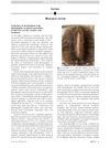
Hair microscopy is a useful and affordable way to diagnose hair disorders.

New imaging tools help doctors better examine hair and scalp health without surgery.
 November 2025 in “Clinical Cosmetic and Investigational Dermatology”
 Digital microscopy can help diagnose hirsutism by measuring hair growth.
 2 citations
,
April 2023 in “Archives of Aesthetic Plastic Surgery”
2 citations
,
April 2023 in “Archives of Aesthetic Plastic Surgery” The phototrichogram is better for measuring hair thickness.
 1 citations
,
March 2014 in “Hair transplant forum international”
1 citations
,
March 2014 in “Hair transplant forum international” Dr. Steve Gabel developed a method for consistent before and after photos in hair transplant surgery using specific camera settings and patient positioning.
1 citations
,
January 2019 in “Indian Journal of Dermatology, Venereology and Leprology”  44 citations
,
January 2019 in “Journal of Translational Medicine”
44 citations
,
January 2019 in “Journal of Translational Medicine” Macrophages are essential for successful skin growth in reconstructive surgery.
 January 2018 in “Communications in computer and information science”
 Researchers developed a computer system to automatically diagnose hair loss by analyzing scalp images.
 14 citations
,
January 2020 in “International Journal of Biological Sciences”
14 citations
,
January 2020 in “International Journal of Biological Sciences” Multiphoton microscopy can effectively assess breast cancer treatment responses without labels.
4 citations
,
October 2024 in “Tissue Engineering and Regenerative Medicine”  24 citations
,
September 2018 in “Lasers in Surgery and Medicine”
24 citations
,
September 2018 in “Lasers in Surgery and Medicine” Multiphoton microscopy can non-invasively tell apart scarring from non-scarring hair loss and could aid in treatment.
3D models from confocal microscopy improve melanoma detection on sun-damaged skin.
 3 citations
,
July 2024 in “Annals of Biomedical Engineering”
3 citations
,
July 2024 in “Annals of Biomedical Engineering” Multiphoton microscopy can effectively detect early endometrial cancer by analyzing collagen changes.
January 2026 in “Updates in clinical dermatology”  8 citations
,
February 2019 in “Scientific Reports”
8 citations
,
February 2019 in “Scientific Reports” Immunofluorescence tomography is a cost-effective method for creating detailed 3-D images of tissues.
56 citations
,
November 2022 in “Biomolecules” Targeting macrophages may improve wound healing.
 6 citations
,
January 2021 in “Journal of The American Academy of Dermatology”
6 citations
,
January 2021 in “Journal of The American Academy of Dermatology” Scalp photography helps patients feel less anxious about hair loss, agree more with doctors on severity, and stay motivated for treatment.
6 citations
,
January 2018 in “Advances in experimental medicine and biology” 13 citations
,
January 2001 in “Skin pharmacology and physiology” Micro-Imager® helps see how drugs spread in human skin.
 2 citations
,
May 2023 in “Experimental dermatology”
2 citations
,
May 2023 in “Experimental dermatology” New imaging techniques can assess and track changes in mouse acne without harm, aiding treatment choices.
119 citations
,
November 1969 in “Journal of Ultrastructure Research” Macrophages help break down collagen around hair follicles during hair growth.
 September 1998 in “Journal of The European Academy of Dermatology and Venereology”
 Phototrichogram helps assess hair loss severity.
March 2026 in “Mendeley Data”