Search
for
Sort by
Research
210-240 / 1000+ results
research Light microscopy of the hair: A simple tool to "untangle" hair disorders
Light microscopy is useful for diagnosing different hair disorders.
research Dosimetry for synchrotron x-ray microbeam radiation therapy
Microbeam radiation therapy's dose distribution changes with depth.
research Importancia del microscopio en el análisis de pelos en la Criminología y en la Criminalística
Microscopes are essential for telling apart human and animal hair in criminal investigations.
research Dermoscopy for the Pediatric Dermatologist Part I: Dermoscopy of Pediatric Infectious and Inflammatory Skin Lesions and Hair Disorders
Dermoscopy improves diagnosis and treatment monitoring for children's skin infections, inflammations, and hair disorders.
research Experimental protocol designed to employ Nd:YAG laser surgery for anterior chamber glaucoma detection via UBM
Targeting the narrowest part of the anterior chamber angle may help prevent pupil block in glaucoma.

research Imaging Tools for Noninvasive Hair Assessment
New imaging tools help doctors better examine hair and scalp health without surgery.
research Detection of Iodine by Scanning Electron Microscopy(SEM) and Energy Dispersive Spectroscopy(EDS) Following Contact with an Iodine Releasing Coating
The method successfully visualizes iodine in biological tissues.
research Presence and future of dermoscopy
Dermoscopy is becoming essential for diagnosing skin conditions and is expected to be a standard tool for dermatologists.
research A single-center, prospective, open-label, pilot study of the safety, local tolerability, and efficacy of ultraviolet-C (UVC) phototherapy for the treatment of great toenail onychomycosis
Trichoscopy is a helpful and quick method to diagnose hair loss without shaving.

research Microscope Matters
The document's conclusion cannot be determined as the content is not available for analysis.

research Multimodal Non-Invasive Skin Imaging in Dermatology: Current Modalities, Clinical Applications, and Future Integration
Integrating various skin imaging technologies improves diagnosis and treatment in dermatology.
research Dissecting Microscope Versus Magnifying Loupes with Transilluntination in the Preparation of Follicular Unit Grafts
Dissecting microscopes give more and better quality hair grafts than magnifying loupes.
research Automated analysis of scanning electron microscopic images for assessment of hair surface damage
A new automated method accurately measures hair damage using microscopic images.
research Electron microscopy in dermatology--basic and clinical research : proceedings of the Joint Meeting for the Japanese Society for Ultrastructural Cutaneous Biology and the Society for Cutaneous Ultrastructure Research, Nara, Japan, 24-27 October 1993
Electron microscopy helps understand skin structure and diseases.

research A 3D microtumour system that faithfully represents ovarian cancer minimal residual disease
The study developed a 3D model that closely imitates remaining ovarian cancer after treatment and identified a potential drug targeting resistant cancer cells.
research 1457 CellutomeTM epidermal harvesting system and in vivo reflectance confocal microscopy as a novel wound healing assessment protocol
The new protocol using Cellutome™ and RCM safely assesses wound healing in detail.
research Clinical Evaluation Including Trichoscopy and Photography
research Scalp Micropigmentation: A Clinicopathologic Correlation
Trichoscopy effectively visualizes scalp micropigmentation without invasive methods.
research 877 Integration of magnetic tweezers and traction force microscopy for exploring the mechanobiology of keratinocyte cell-cell and cell-matrix anchoring junctions
The conclusion is that a new method combining magnetic tweezers and traction force microscopy may help understand skin cell interactions and diseases.
research Scalable fabrication, compartmentalization and applications of living microtissues
Continuous microfluidic processes can help scale up microtissue production for industrial and clinical use.
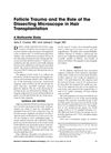
research Follicle trauma and the role of the dissecting microscope in hair transplantation
Using a microscope during hair transplants cuts damage to follicles in half and could improve hair growth.
research Laser capture microdissection as a method for investigating the human hair follicle microbiome reveals region-specific differences in the bacteriome profile
Laser-capture microdissection effectively analyzes hair follicle microbiomes, revealing region-specific bacterial differences.
research Dynamic Trichoscopy
Trichoscopy is useful for diagnosing and monitoring hair and scalp conditions over time.
research Visual imaging of calcium ion distribution in acetone and tape stripping damaged canine epidermis.
The method visualized calcium ions in damaged canine skin, showing potential for studying skin recovery.
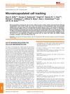
research Microencapsulated cell tracking
Microencapsulation helps protect and track therapeutic cells, showing promise for treating various diseases, but more work is needed to improve the technology.

research OPTICAL COHERENCE TOMOGRAPHY AS AN ADVANCED TOOL IN DIFFERENT APPLICATIONS
Optical Coherence Tomography has potential in diagnosing hair loss and monitoring blood clotting, and could be improved for deeper tissue observation and better hair loss understanding.
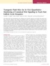
research Transgenic Flash Mice for In Vivo Quantitative Monitoring of Canonical Wnt Signaling to Track Hair Follicle Cycle Dynamics
Researchers developed a mouse model that tracks hair growth using bioluminescence, improving accuracy in studying hair cycles.
research Review paper The use of reflectance confocal microscopy in selected inflammatory skin diseases
Reflectance confocal microscopy is a useful, non-invasive tool for diagnosing some skin diseases, with potential for future improvements.
research Microimaging of hairless rat skin by magnetic resonance at 900 MHz
MRI can effectively image skin structures noninvasively.